斑塊分析
2017-04-08 10:03:16 by admin ![]() 14754
14754

斑塊視界原創(chuàng)

核磁共振動(dòng)脈硬化斑塊分析系統(tǒng)MRI-PlaqueView是基于核磁共振影像的斑塊成像斑塊分析軟件產(chǎn)品。
01/斑塊分析
該產(chǎn)品已獲得美國藥監(jiān)局批準(zhǔn)用于頸動(dòng)脈疾病的臨床,該產(chǎn)品也可用于主動(dòng)脈及周動(dòng)脈的斑塊分析研究。
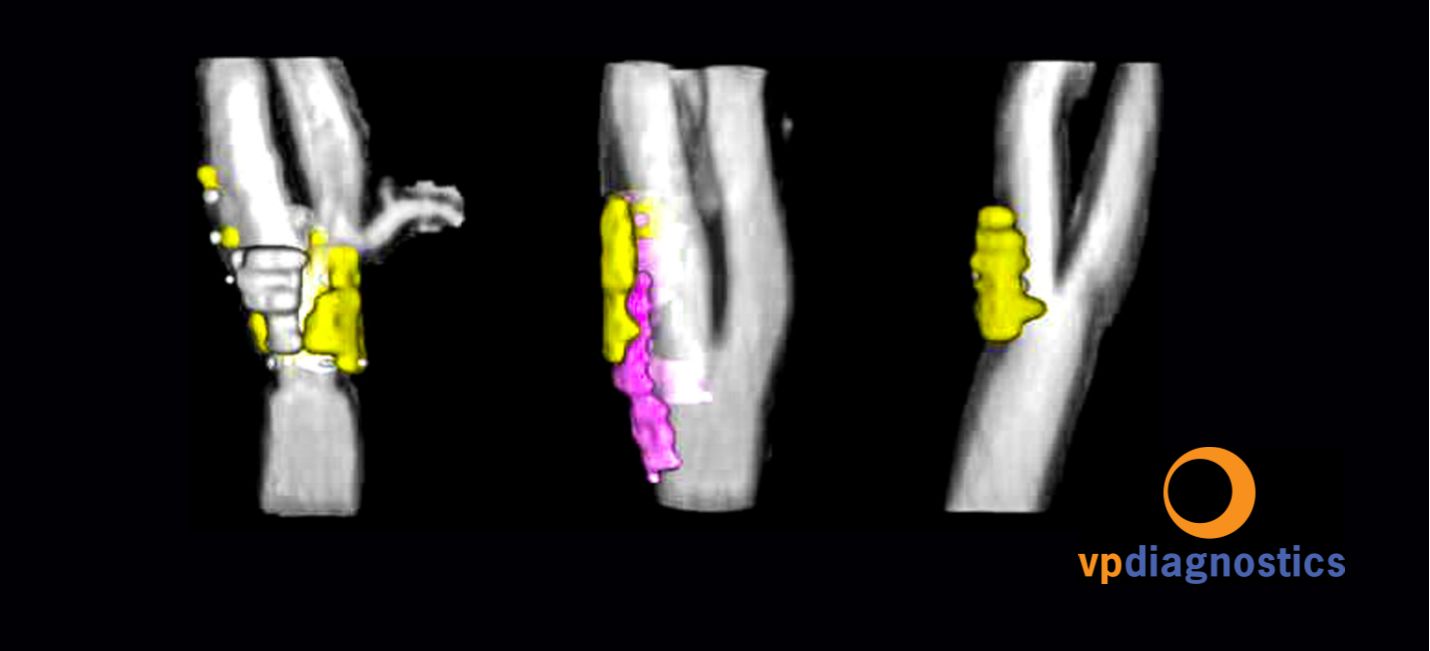
該產(chǎn)品可以快捷地提供動(dòng)脈硬化疾病的分析,提供斑塊的形態(tài)及成分的定量檢測(cè)及其三維立體分布。

MRI-PlaqueView提供自動(dòng)、優(yōu)化的操作流程,引導(dǎo)幫助用戶進(jìn)行斑塊分析。幾分鐘之內(nèi),即可以完成血管腔、血管壁及內(nèi)部形態(tài)及成分(如軟斑塊,鈣化斑塊,斑塊內(nèi)出血)的分析,分析包括面積,厚度,距離以及血管狹窄程度,并可以存錄。

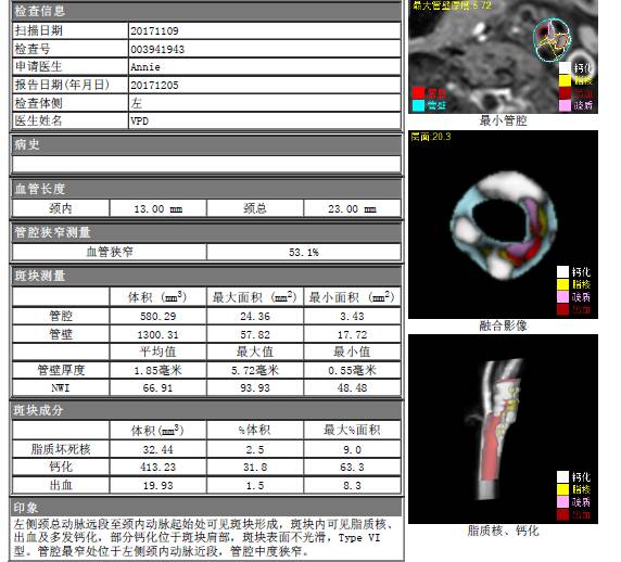
MR-VPD分析報(bào)告樣本:從“形態(tài)分布”到“數(shù)據(jù)信息”,全方位分析斑塊
評(píng)估報(bào)告匯集斑塊評(píng)估分析的重要發(fā)現(xiàn),明了全面,有利于血管外科醫(yī)生、神經(jīng)科醫(yī)生、介入治療醫(yī)生、以及心腦血管醫(yī)生對(duì)病人的干預(yù)防治。
02/獲取方法
產(chǎn)品購買信息:核磁共振動(dòng)脈硬化斑塊分析系統(tǒng)MRI-PlaqueView目前提供四種版本供不同用戶選擇,包括:斑塊負(fù)荷分析,斑塊內(nèi)出血分析,斑塊成分分析和自動(dòng)斑塊分析。
軟件的授權(quán)可以按使用次數(shù)或者無限量形式購買。進(jìn)一步信息請(qǐng)下載 核磁共振動(dòng)脈硬化分析系統(tǒng)-MRI-PlaqueView介紹 或者直接 聯(lián)系我們 以安排咨詢演示試用。
斑塊成像及基于MRI-PlaqueViewTM 的斑塊分析與MRI設(shè)備生產(chǎn)商GE,Philips 具有全球戰(zhàn)略合作關(guān)系上:
03/科研基礎(chǔ)
美國衛(wèi)生部保健研究及質(zhì)量局(The Agency for Healthcare Research and Quality (AHRQ)of U.S. Department of Health and Human Services (HHS))2010年8月發(fā)布易損斑塊技術(shù)概述(Technical Brief: Vulnerable Atherosclerotic Plaque)。我公司的核磁共振動(dòng)脈硬化斑塊分析系統(tǒng)-MRI-PlaqueView產(chǎn)品收錄在第60 頁。
我公司正在進(jìn)行由美國國立衛(wèi)生研究院(NIH – National Institutes of Health)資助的多中心、前瞻性頸動(dòng)脈斑塊評(píng)估臨床研究。
這項(xiàng)臨床研究的目的是驗(yàn)證基于核磁共振動(dòng)脈硬化斑塊分析系統(tǒng)的斑塊分析診斷技術(shù)對(duì)中風(fēng)及相關(guān)疾病的預(yù)警能力。
這一臨床在市場(chǎng)研究報(bào)告(MarketResearch.com)2010年4季度發(fā)布的全球動(dòng)脈硬化臨床試驗(yàn)回顧 (Arteriosclerosis Global Clinical Trials Review, Q4, 2010) 中收錄。
04/ 驗(yàn)證文獻(xiàn):
核磁共振動(dòng)脈硬化斑塊分析系統(tǒng)使用的核心算法技術(shù)的準(zhǔn)確性、可重復(fù)性以及有效性已經(jīng)在50多篇專業(yè)論文中提供了充分的驗(yàn)證。以下是摘選的相關(guān)文獻(xiàn)。
1.由VPDiagnostics協(xié)助的研究
Automated in vivo segmentation of carotid plaque MRI with Morphology-Enhanced probability maps. (Liu, Magn Reson Med. 2006)
Automated measurement of mean wall thickness in the common carotid artery by MRI: a comparison to intima-media thickness by B-mode ultrasound. (Underhill, J Magn Reson Imaging. 2006)
Magnetic resonance imaging of carotid atherosclerosis: plaque analysis. (Kerwin, Top Magn Reson Imaging. 2007)
Signal features of the atherosclerotic plaque at 3.0 Tesla versus 1.5 Tesla: impact on automatic classification. (Kerwin,J Magn Reson Imaging. 2008) PDF
2.涉及核磁共振動(dòng)脈硬化分析系統(tǒng)-MRI-PLAQUEVIEW的技術(shù)的相關(guān)研究
Scan-rescan reproducibility of carotid atherosclerotic plaque morphology and tissue composition measurements using multicontrast MRI at 3T. (Li, J Magn Reson Imaging. 2010)
Carotid intraplaque hemorrhage imaging at 3.0-T MR imaging: comparison of the diagnostic performance of three T1-weighted sequences. (Ota, Radiology. 2010)
Carotid plaque morphology and composition: initial comparison between 1.5- and 3.0-T magnetic field strengths. (Underhill, Radiology. 2008) PDF
Reader and platform reproducibility for quantitative assessment of carotid atherosclerotic plaque using 1.5T Siemens, Philips, and General Electric scanners. (Saam, JMRI 2007)
Intra- and interreader reproducibility of magnetic resonance imaging for quantifying the lipid-rich necrotic core is improved with gadolinium contrast enhancement. (Takaya, JMRI 2007)
Quantitative evaluation of carotid plaque composition by in vivo MRI. (Saam, ATVB 2005) PDF
In vivo quantitative measurement of intact fibrous cap and lipid-rich necrotic core size in atherosclerotic carotid plaque: comparison of high-resolution, contrast-enhanced magnetic resonance imaging and histology. (Cai, Circulation 2005) PDF

 京公網(wǎng)安備 11010502042883號(hào)